Services and Patient Information

X-Rays

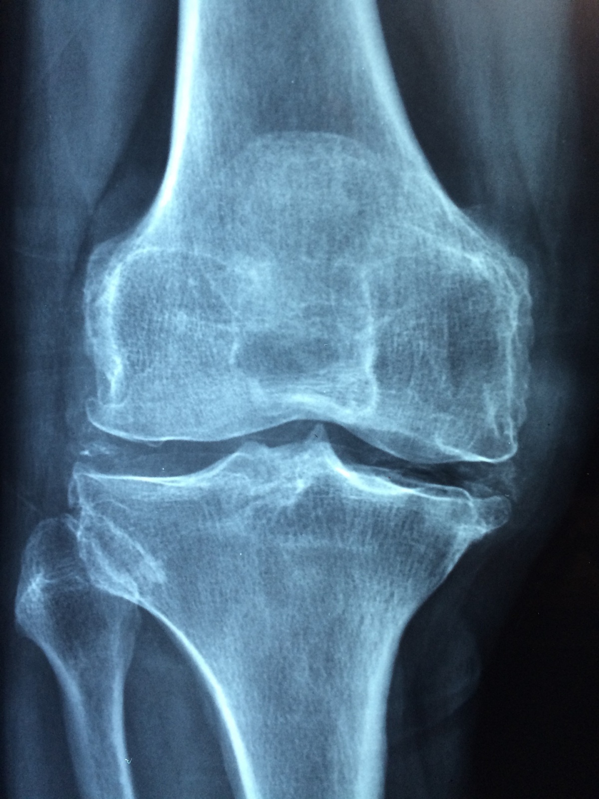
X-Rays are a quick and painless way for a physician to look at and evaluate the bones of your body.

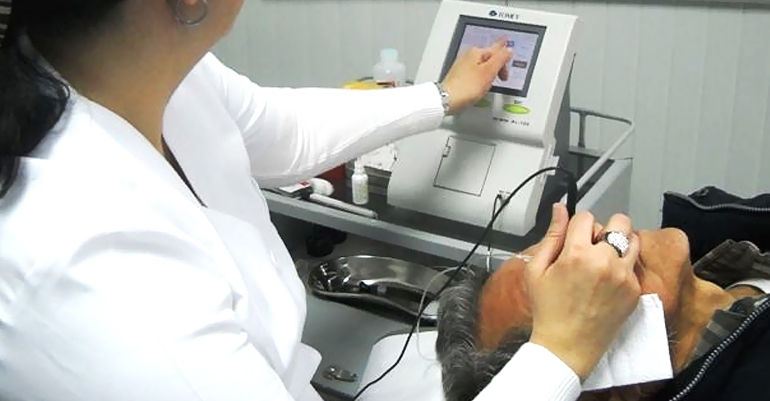
Our experienced technologists are friendly and efficient. The exam is completed quickly. Your images will be reviewed by the Radiologist, who will report the findings to your referring physician. You can contact your physician for the results.

How the X-Ray is Captured

X-ray beams pass through your body and are absorbed in different amounts depending on the density of the material they pass through. The resulting shadows and reflections are digitally captured on a computer screen. Dense materials, such as bone and metal, show up as white on X-rays. The air in your lungs shows up as black. Fat and muscle appear as varying shades of gray.

Patient Preparation

  • When you come in for an X-ray, depending on which body part is being imaged, you may be asked to remove your jewelry or garments with metal closures. These items can block part of the image.
  • You will be asked to either lie on a table, sit or stand and a lead apron may be draped over part of your body to shield it from the X-rays.
  • We are sensitive to your individual needs and will help you in any way we can to make you feel as comfortable as possible.

Your X-rays will be reviewed by the Radiologist, who will report the findings to your referring physician. You can contact your physician for the results.

X-Ray service is provided on a first come, first served, walk-in basis.

Erie St Clair Radiology

Ultrasound

An ultrasound scan is a safe, painless and effective medical procedure that uses high frequency sound waves to create an image of the internal organs such as the stomach, liver, heart, tendons, muscles, joints and blood vessels. Obstetric ultrasounds are done to check and monitor the growth of the fetus in a mother’s womb. Our staff are sensitive to your needs and it’s our goal to make this procedure as comfortable as possible.

Your ultrasound images will be reviewed by the radiologist, who will report the findings to your referring physician. You can contact your physician for the results.

How the Ultrasound is Captured

Ultrasound uses high frequency sound waves to create images.

During the ultrasound, the skin over the examination area is coated with a wet gel to allow for smooth movement of the wand (transducer) over the skin. Positioning the transducer at certain locations and angles, creates waves, or echoes, which are then analyzed by a sophisticated computer to create real-time, multi-dimensional digital electronic images.

Patient Preparation

  • Wear comfortable, loose-fitting clothing for your ultrasound exam. You will need to remove all clothing and jewelry in the area to be examined. You may be asked to wear a gown during the procedure. Our staff will guide you through the process and be sensitive to your needs.
  • Depending on the type of examination, you may be instructed not to eat or drink for as many as 8 hours before your appointment. You may be asked to finish drinking 5 glasses of water an hour prior to your exam so that your bladder is full when the scan begins. However, if this is the case, you will be advised (upon booking) so you will be ready for your procedure.
  • During the ultrasound, a clear gel is applied to the area of the body being examined. The ultrasound tech will then use the wand (transducer) against the skin and sweep over the area.
  • Most ultrasound examinations are completed within 30 minutes or less.

Your ultrasound images will be reviewed by the radiologist, who will report the findings to your physician within 1 business day. Your physician will then advise you of the results and discuss them with you.

Bone Mineral Density

Bone density testing (via DEXA) determines if you have osteoporosis, a disease that causes bones to become more fragile and more likely to break in the next few years.

DEXA scan is a simple, quick and non-invasive procedure that uses very small amounts of X-ray to measure calcium content of the bone and to detect the presence of weakened bone density and other bone minerals which are packed into a segment of bone. The bones that are most commonly tested are in the spine, hip and forearm.

Your BMD images will be reviewed by the Radiologist, who will report the findings to your Physician. Your Physician will then advise you of your results and discuss them with you.

In general, people with a bone mineral density significantly lower than normal for their age and sex are more likely to break a bone. It is recommended that patients with a family history or who are at risk for osteoporosis, get a bone density test.

How to Prepare

  • Don’t take calcium or iron supplements for 24 hours before the exam.
  • If you’ve had an injection of barium or contrast dye for a CT scan or MRI, wait 7 days before having a BMD DEXA. The contrast dye could interfere with your bone density test.
  • Avoid wearing clothes with metal zippers, belts, or buttons.

There is very little risk to taking the test. You’re exposed to very low levels of radiation, less than a chest X-ray or an airplane flight.

Opthalmology

There are two types of eye ultrasounds which may be ordered by your Ophthalmologist. An A-scan takes measurements of the eye, and a B-scan helps to see the structures in the back of the eye.

No preparation is needed for the tests.  Do not wear eye make-up to the tests.

There is no pain with the test, as anesthetic drops are used to temporarily numb your eye, which typically lasts for about 30 minutes, and does not affect your vision.  After the test, do not rub your eyes until the anesthetic has completely worn off. This is to protect you from accidentally injuring your eye.

A-scan (Biometry)

An A-scan ultrasound is used to measure the length of the eye to determine the correct lens implant for cataract surgery. The test will typically take about 30 minutes.  Both eyes are scanned for comparison (even if you have had a previous cataract surgery).

You will be lying down for this test. The test is performed with the eye open. After the anesthetic drop is applied, a small plastic cup will be placed on the surface of your eye and filled with fluid (saline). A small pencil sized probe is then placed into the water to perform the measurements.

B-scan (B-mode)

A B-scan ultrasound is used to image the structures within the eye when there is an injury, to monitor an existing condition, or to image the eye after a surgery.

The B-scan may help in the diagnosis of tumors, foreign objects, bleeding within the clear vitreous gel that fills the back of the eye, detachments of the membranes, and conditions involving the optic nerve.

You will be lying down for this test which will typically take about 20-30 minutes.  After the anesthetic drop is applied, a small soft probe about the size of a highlighter is applied to the eye, and you will be asked to look in different directions while images are taken.  To help provide the best images, the eye is kept open (as eyelids may decrease image quality) and a special gel is used that is PH balanced for the eye.  There is no pain with the test, although some people may experience an odd sensation when the cool gel touches the eye. Upon completing the test, the eye is rinsed with fluid (saline) to remove any remaining gel.

UBM

A specialized high resolution B-scan ultrasound may also be ordered by your doctor to help image the structures at the front of your eye such as the cornea and iris. This test is performed in the same manner as the B-scan, but uses a special soft covering for the probe that is filled with fluid (saline).

How Should I Prepare?

  • Bring your OHIP card and doctor’s requisition.
  • Arrive 15 minutes early. Sometimes services take longer than expected.
  • If you can’t make your appointment or are late, please call us. We may have to rebook it.
  • Take your medications as per your doctor’s orders.
  • Avoid perfumes and fragrances as a courtesy to others
  • Avoid clothing with metal buttons, buckles, or zippers.
  • Tell your technologist if you are or could be pregnant.
  • Diabetics with ‘clear liquid’ orders should schedule in the morning. Discuss insulin dosages with your doctor.
  • Please refer to the back of the requisition for exams that require prior preparation.

For detailed information, download our printable one-page exam prep document.